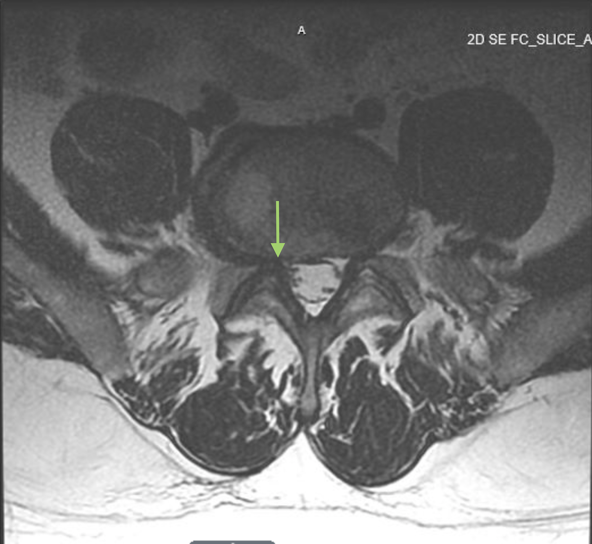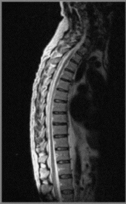

( B) Sagittal T2-weighted image shows well-hydrated intervertebral disc spaces. ( A) Sagittal T1-weighted image shows normal marrow signal, alignment, and vertebral body heights. ( B) Axial GRE image shows normal canal and neural foramen.įigure 14-2. ( A) Sagittal fat-saturated T2-weighted image shows well-hydrated intervertebral disc spaces and normal cervical cord signal. Gradient-recalled echo (GRE) sequences are helpful for detecting blood products associated with cord hemorrhage in the setting of trauma.įigure 14-1. Proton density (PD) sequences may be useful in detecting cord signal abnormalities associated with demyelinating diseases such as multiple sclerosis (MS).

Contrast-enhanced T1-weighted sequences are helpful for evaluation of suspected neoplasm, infection, and inflammatory diseases. The intervertebral disc is normally high signal intensity on T2-weighted images because of its high water content but frequently loses signal over time with loss of water content. In the adult, the normal vertebral marrow generally has intermediate to high signal intensity on the T1-weighted images and low signal intensity on T2-weighted images. Short tau inversion recovery (STIR) or fat-saturated T2-weighted sequences are also invaluable for increased sensitivity of marrow edema. Additional coronal images may be helpful especially in the setting of scoliosis. The standard spine MRI protocol includes imaging in the sagittal and axial planes using T1- and T2-weighted sequences ( Figures 14-1 and 14-2). Intervertebral discs separate the vertebrae and serve as shock absorbers. The posterior elements include the articulating processes, lamina, and spinous process. Except for the first and the second cervical vertebrae, the vertebrae share a similar structure including a vertebral body containing trabecular bone. The spine is composed of seven cervical, twelve thoracic, and five lumbar vertebrae as well as the fused sacrum and coccyx vertebral elements. The spine is composed of multiple vertebrae, which protect the spinal cord and proximal spinal nerves. Given its complex anatomy and length, the spine remains one of the most difficult parts of the skeletal system to evaluate. As the spinal cord has limited healing ability, an acute myelopathy is an emergency and should prompt urgent imaging, preferably with MRI given its superior evaluation of the spinal cord and canal. Classic symptoms of myelopathy include bladder and bowel incontinence, spasticity, weakness, and ataxia. However, myelopathy is caused by mechanical spinal cord compression or by intrinsic lesions of the spinal cord. This results in specific sensory deficits and muscle group weakness. Radiculopathy results from mechanical compression or irritation of a spinal nerve, often within a lateral recess or neural foramina. Patients with spine disorders may also present with radiculopathy and myelopathy. Fever or history of malignancy should raise suspicion and urgency.

While back pain is epidemic and associated with great disability, back pain without neurologic compromise is usually not an emergency. When considering performing and interpreting imaging of the spine, it is important to first understand the clinical context. Although diseases of the spine are very common, clinical syndromes may mimic each other, necessitating imaging such as MRI for diagnosis and patient management.

Magnetic resonance imaging (MRI) has become the examination of choice for imaging the spine and its contents.


 0 kommentar(er)
